Saturday, 6 February 2016

Ocular myasthenia gravis


Ocular myasthenia gravis

Ocular myasthenia gravis (MG) is a disease of the neuromuscular junction resulting in hallmark variability in muscle weakness and fatigability. MG is an autoimmune disease where anomalous antibodies are produced against the naturally occurring acetylcholine receptors in voluntary muscles. MG is an autoimmune disease where anomalous antibodies are produced against the naturally occurring acetylcholine receptors in voluntary muscles. MG may be limited to the muscles of the eye (ocular MG), leading to abrupt onset of weakness/fatigability of the eyelids or eye movement. MG may also involve other muscle groups.
The most common symptoms seen in patients with ocular MG are diplopia (double vision), ptosis (droopy eyelids), and incomplete eye closure. Compared to other involved skeletal muscles, only slight weakness of the extra ocular muscle may cause diplopia and visual disturbances to occur. These symptoms occur due to weakness of the muscle that control eyeball and eyelid movement. Light sensitivity due to sluggish pupils may occur in some patients. Symptoms are frequently influenced by environmental, emotional, and physical factors. Some of these factors include bright sunlight, extreme temperature, emotional stress, illness, surgery, menstruation, and pregnancy, among others. Symptoms tend to be worse at the end of the day.

Symptoms

 Diplopia or double vision results when the eyes cannot be focused as desired due to weakness of one or more of the extraocular muscles which control eye movement. This most often occurs when looking up or to the side. To compensate for the weakness, the patient may tilt his/her head or turn their face to allow the stronger eye to work. For example, if the muscle which allows the eye to look upward is weak, the patient could tilt their head back to look up.
 Ptosis is the drooping of one or both eyelids, also due to muscle weakness. Fluttering or twitching of the eyelid may occasionally be seen. If both eyelids are droopy, one may be worse than the other.
Nystagmus  or constant involuntary repeated movement of the eyeball in any direction may also occur in one or both eyes.

Treatment


Treatment of ocular MG is aimed at relieving the symptoms of ptosis and diplopia, as well as preventing the development of generalized MG symptoms. Immune suppression with steroids is often the main therapy. Steroid doses must be increased slowly because of a risk of precipitating myasthenic crisis. After achieving the highest target dose, steroids are then slowly tapered down to the lowest effective dose. Often, acetylcholinesterase inhibitors such as pyridostigmine and neostigmine are also employed to help control symptoms. When steroids are contraindicated, acetylcholinesterase inhibitors can be tried as the primary therapy. Steroid-sparing agents such as azathioprine and mycophenolate may also have a role in treating ocular MG. Other treatments for MG include plasmapheresis, intravenous immunoglobulin, and other immunosuppressive agents, but these are rarely required for ocular MG. Patients should also be evaluated for thymoma. Thymoma should be resected surgically. Ocular MG without thymoma is not usually treated with thymectomy. Topical agents may be useful as additional therapy for mild or moderate ptosis. Nonpharmacologic treatments include occlusive devices, prisms, eyelid supports, contact lenses, and (in long-standing, stable cases) strabismus surgery or eyelid elevation surgery.

Monday, 1 February 2016

Guillain-Barre Syndrome


Guillain-Barre Syndrome

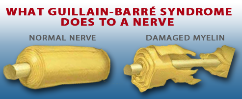
Guillain-Barre syndrome is a rare disorder in which your body's immune system attacks your nerves. Weakness and tingling in your extremities are usually the first symptoms.
These sensations can quickly spread, eventually paralyzing your whole body. In its most severe form Guillain-Barre syndrome is a medical emergency. Most people with the condition must be hospitalized to receive treatment

Symptoms
Guillain-Barre syndrome often begins with tingling and weakness starting in your feet and legs and spreading to your upper body and arms. In about 10 percent of people with the disorder, symptoms begin in the arms or face. As Guillain-Barre syndrome progresses, muscle weakness can evolve into paralysis.
Signs and symptoms of Guillain-Barre syndrome may include:
  • Sensations in your fingers, toes, ankles or wrists
  • Weakness in your legs that spreads to your upper body
  • Unsteady walking or inability to walk or climb stairs
  • Difficulty with eye or facial movements, including speaking, chewing or swallowing
  • Severe pain that may feel achy or cramp-like and may be worse at night
  • Difficulty with bladder control or bowel function
  • Rapid heart rate
  • Low or high blood pressure
  • Difficulty breathing


Causes
The exact cause of Guillain-Barre syndrome isn't known. The disorder usually appears days or weeks after a respiratory or digestive tract infection. Rarely, recent surgery or immunization can trigger Guillain-Barre syndrome.
Guillain-Barre syndrome can affect all age groups. But you're at slightly greater risk if:
  • You're a man
  • You're an older adul 
Guillain-Barre syndrome may be triggered by:
  • Infection with campylobacter, a type of bacteria often found in undercooked poultry
  • Influenza virus
  • Epstein-Barr virus
  • HIV, the virus that causes AIDS
  • Mycoplasma pneumonia
  • Surgery
  • Hodgkin's lymphoma
  • Rarely, influenza vaccinations or childhood vaccinations

Complications
Guillain-Barre syndrome affects your nerves. Because nerves control your movements and body functions, people with Guillain-Barre may experience:
  • Breathing difficulties.
  • Residual numbness or other sensations.
  • Heart and blood pressure problems
  • Heart and blood pressure problems
  • Bowel and bladder function problems.
  • Blood clots.
  • Pressure sores
Severe, early symptoms of Guillain-Barre syndrome significantly increase the risk of serious long-term complications. Rarely, death may occur from complications such as respiratory distress syndrome and heart attack.
Tests and diagnosis
Guillain-Barre syndrome can be difficult to diagnose in its earliest stages. Its signs and symptoms are similar to those of other neurological disorders and may vary from person to person.
  • Spinal tap (lumbar puncture)
  • Electromyography
  • Nerve conduction studies.

Treatments 

There's no cure for Guillain-Barre syndrome. But two types of treatments can speed recovery and reduce the severity of the illness:


Saturday, 30 January 2016

Risk Factors


What are the risk factors for brain and spinal cord tumors?

A risk factor is anything that affects your chance of getting a disease such as a brain or spinal cord tumor. Different types of cancer have different risk factors. Some risk factors, like smoking, you can change. Others, like your age or family history, can’t be changed.
Most brain tumors are not linked with any known risk factors and have no obvious cause. But there are a few factors that can raise the risk of brain tumors.

Radiation exposure
The best known environmental risk factor for brain tumors is radiation exposure, most often from radiation therapy to treat some other condition.

Family history
Most people with brain tumors do not have a family history of the disease, but in rare cases brain and spinal cord cancers run in families. In general, patients with familial cancer syndromes tend to have many tumors that first occur when they are young.
                   

Neurofibromatosis type 1 (NF1)
This genetic disorder, also known as von Recklinghausen disease, is the most common syndrome
linked to brain or spinal cord tumors. People with this condition have higher risks of schwannomas, meningiomas, and certain types of gliomas, as well as neurofibromas (benign tumors of peripheral nerves). Changes in the NF1 gene cause this disorder. These changes are inherited from a parent in about half of all cases. In the other half, the NF1 gene changes occur before birth in people whose parents did not have this condition.

Neurofibromatosis type 2 (NF2)
This condition, which is much less common than NF1, is associated with vestibular schwannomas (acoustic neuromas), which almost always occur on both sides of the head. It is also linked with an increased risk of meningiomas or spinal cord ependymomas. Changes in the NF2 gene are responsible for neurofibromatosis type 2. Like NF1, the gene changes are inherited in about half of cases or may occur before birth in children without a family history.

Tuberous sclerosis
People with this condition may have subependymal giant cell astrocytomas (SEGAs), which are low-grade astrocytomas that develop beneath the ependymal cells of the ventricles). They may also have other benign tumors of the brain, skin, heart, kidneys, and other organs. This condition is caused by changes in either the TSC1 or theTSC2 gene. These gene changes can be inherited from a parent, but most often they develop in people without a family history.

Von Hippel-Lindau disease
People with this condition tend to develop benign or cancerous tumors in different parts of the body, including hemangioblastomas (blood vessel tumors) in the brain, spinal cord, or retina, as well as tumors of the inner ear, kidney, adrenal gland, and pancreas. It is caused by changes in the VHL gene. Most often the gene changes are inherited, but in some cases the changes happen before birth in people whose parents don’t have them.

Li-Fraumeni syndrome
People with this condition are at higher risk for developing gliomas, along with breast cancer, soft tissue sarcomas,leukemia, and adrenal gland cancer, and certain other types of cancer. It is caused by changes in the TP53 gene.

Other syndromes
Other inherited conditions are also linked with increased risks of certain types of brain and spinal cord tumors, including:
·                         Gorlin syndrome (basal cell nevus syndrome)
·                         Turcot syndrome
·                         Cowden syndrome
Some families may have genetic disorders that are not well recognized or that may even be unique to a particular family.

Immune system disorders
People with impaired immune systems have an increased risk of developing lymphomas of the brain or spinal cord (known as primary CNS lymphomas). Lymphomas are cancers of lymphocytes, a type of white blood cell that fights disease. Primary CNS lymphoma is less common than lymphoma that develops outside the brain.
A weakened immune system can be congenital (present at birth), or it can be caused by treatments for other cancers, treatment to prevent rejection of transplanted organs, or diseases such as the acquired immunodeficiency syndrome (AIDS).

Unproven effects on brain tumor risk
·         Cell phone use
·         microwave ovens
·         Radar
·         Satellite stations
·         Vinyl chloride (a chemical used to manufacture plastics)
·         Petroleum products
·         Exposure to aspartame (a sugar substitute),
·         Exposure to electromagnetic fields
·         Infection with certain viruses


Thursday, 21 January 2016

Glioma

Glioma

Glioma is a type of tumor that occurs in the brain and spinal cord. Gliomas begin in the gluey supportive cells (glial cells) that surround nerve cells and help them function. “Glioma” is a general term used to describe any tumor that arises from the supportive (“gluey”) tissue of the brain. This tissue, called “glia,” helps to keep the neurons in place and functioning well.

Three types of glial cells can produce tumors. Gliomas are classified according to the type of glial cell involved in the tumor.

There are 3 main types of glioma                                                                     
·     Astrocytoma
·     Oligodendroglioma
·     Ependymoma
An astrocyte will produce astrocytomas (including glioblastomas), an oligodendrocyte will produce oligodendrogliomas, and ependymomas come from ependymal cells. Tumors that display a mixture of these different cells are called mixed gliomas.

Astrocytomas (including glioblastoma multiforme)
Astrocytomas are the most common type of glioma in both adults and children. They develop from cells called astrocytes. The astrocytes are the cells of the brain that support the nerve cells (neurones). Astrocytomas can be slow growing (low grade) or fast growing (high grade). Some are very localised (focal). This means it is easy to see the border between the tumour and normal brain tissue on a scan or during an operation. Focal astrocytomas are more often diagnosed in children and are not common in adults
.
Other astrocytomas are called diffuse. These do not have a clear boundary between the tumour and normal brain tissue.

Anaplastic astrocytoma (also called grade 3 astrocytoma) and glioblastoma multiforme (GBM or grade 4 astrocytoma) are the most common type of brain tumour in adults. These are malignant (high grade) brain gliomas. They can sometimes spread to other parts of the brain.

Oligodendroglioma
An oligodendroglioma tumor is a slow-growing brain tumor that usually occurs in young adults. These tumors are frequently located within the frontal, temporal or parietal lobes and cause seizures in a relatively high percentage of patients. Many oligodendrogliomas contain little specks of calcium (bone) and can easily bleed.

On imaging these tumour commonly present as a round or oval sharply marginated mass involving the cortex or subcortical white matter, with low attenuation on CT, hypointense compared to gray matter on T1 and hyperintense compared to gray matter on T2-weighted MRI images. The attenuation or signal can be eventually heterogeneous due calcification, cystic degeneration and hemorrhage.

Ependymomas
About 2 out of every 100 brain tumours (2%) are ependymomas. These develop from cells called ependymal cells. These cells line the fluid filled areas of the brain (the ventricles) and the spinal cord. Their job is to repair any damaged nerve tissue.

Most ependymomas are diagnosed in children or young adults and can occur in any part of the brain or spinal cord. In older patients they tend to occur in the lower part of the spinal cord. Ependymomas can be high or low grade, but the cells' appearance under a microscope does not always fit with their behaviour. So the grade may not tell you much about how likely the tumour is to grow or spread.
Sometimes ependymomas can spread to other parts of the central nervous system, through the fluid that circulates around the brain and spinal cord.


Treatment for Gliomas
The best treatment for an individual patient takes into account the tumor location, potential symptoms, and potential benefits versus risks of the different treatment options (modalities).
Treatment for a glioma is customized to the individual patient and may include surgery, radiation therapy, chemotherapy, or observation. Treatment for patients with brain tumors is best done by a multi-disciplinary team. This includes neurosurgeons, medical neuro-oncologists and radiation therapists


Friday, 15 January 2016

Brain (cerebral) aneurysm


Brain (cerebral) aneurysm

A brain (cerebral) aneurysm is a bulging, weak area in the wall of an artery that supplies blood to the brain. In most cases, a brain aneurysm causes no symptoms and goes unnoticed. In rare cases, the brain aneurysm ruptures, releasing blood into the skull and causing a stroke.


When a brain aneurysm ruptures, the result is called a subarachnoid hemorrhage. Depending on the severity of the hemorrhage, brain damage or death may result.

The most common location for brain aneurysms is in the network of blood vessels at the base of the brain called the circle of Willis.

Causes
Ø  Hardening of the arteries (atherosclerosis)
Ø  Aging
Ø  Family history
Ø  Previous aneurysm
Ø  Gender.
Ø  Race
Ø  High blood pressure
Ø  Smoking
Symptoms

v  A sudden, severe headache that is different from past headaches.
v  Neck pain.
v  Nausea and vomiting.
v  Sensitivity to light.
v  Fainting or loss of consciousness.
v  Seizures.

Tests

you have a brain aneurysm, you may have the following tests:
  • Computed tomography (CT) scan.
  • Computed tomography angiogram (CTA) scan.
  • Magnetic resonance angiography (MRA)
  • Cerebral angiogram.

Treatment

The following surgeries are used to treat both ruptured and unruptured brain aneurysms:

  • Embolization
  • Surgical clipping.

Wednesday, 13 January 2016

Seizure


Seizure

A seizure is when an electrical discharge happens suddenly in your brain. This occurs from a problem in brain cells that can lead to changes in what you feel and how you act. It can also cause symptoms like muscle spasms, limb twitches, and loss of consciousness.


A seizure refers to a one-time event. If you have more than one seizure, then your doctor may diagnose the condition as a larger disorder. The Epilepsy Researchers reports that once you have a single seizure with no obvious cause, then you have about a 50 percent chance of having another seizure. The second seizure often occurs within half a year of the first one.

Causes

Under normal circumstances, your brain cells use electrical activity to communicate through neurons transmitting information. Seizures happen when brain cells behave abnormally, causing neurons to misfire and send the wrong signals.


According to the Researchers, the cause of this abnormal brain cell response is unclear in about 50 percent of people who have a seizure. But certain conditions may lead to seizures. These include:
·         Alzheimer’s disease/dementia
·         heart problems like stroke or heart attack
·         injury to your head or brain (including before birth)
·         lupus
·         meningitis

Researchers believe that certain genes can also cause seizures, as well as some conditions that you may have from birth.

Types


Seizures can be classified into two primary types: partial and generalized.
Partial seizures:  Partial seizures begin in a limited part of your brain. If they originate on one side of your brain and spread to other areas, they are called simple partial seizures. If they begin in an area of your brain that affects consciousness, they are called complex partial seizures.
Generalized seizures:  Generalized seizures begin on both sides of your brain at the same time. Because they spread quickly, it can be impossible to tell where they’ve originated. This makes certain kinds of treatments more difficult.

Treatments

·         Medicines
·         Surgery

·         Diet Changes

Blood pressure / Hypertension


Blood pressure

Blood pressure is the force exerted by the blood against the walls of blood vessels, and the magnitude of this force depends on the cardiac output and the resistance of the blood vessels.


Hypertension is having a blood pressure higher than 140 over 90 mmHg, a definition shared by all the medical guidelines. This means the systolic reading (the pressure as the heart pumps blood around the body) is over 140 mmHg (millimeters of mercury) or the diastolic reading (as the heart relaxes and refills with blood) is over 90 mmHg.Based on American Heart Association (AHA),

1. Normal blood pressure is below 120 systolic and below 80 diastolic
2. Prehypertension is 120-139 systolic or 80-89 diastolic
3. Stage 1 high blood pressure (hypertension) is 140-159 systolic or 90-99 diastolic
4. Stage 2 high blood pressure (hypertension) is 160 or higher systolic or 100 or higher diastolic
5. Hypertensive crisis (a medical emergency) is when blood pressure is above 180 systolic or above 110 diastolic.

Symptoms
1. headaches
2. shortness of breath or nosebleeds                                                


Causes of hypertension
1. Physical inactivity
2. A salt-rich diet through processed and fatty foods
3. Alcohol and tobacco use.
4. Age
5. Size

6. Lifestyle

Tests

To measure your blood pressure, your doctor or a specialist will usually place an inflatable arm cuff around your arm and measure your blood pressure using a pressure-measuring gauge.


Treatments

Changing your lifestyle can go a long way toward controlling high blood pressure. Your doctor may recommend you eat a healthy diet with less salt, exercise regularly, quit smoking and maintain a healthy weight.
In addition to diet and exercise, your doctor may recommend medication to lower your blood pressure.
Your blood pressure treatment goal depends on how healthy you are.